
Krembil’s annual Research Day offers trainees, investigators, and staff a valuable opportunity to network and celebrate research achievements. This year, the Krembil Research Institute (Krembil) celebrated 25 years of this annual event.
On Thursday, April 24, 2025, more than 200 trainees, faculty, and staff gathered at the MaRS Centre. The day provided the Krembil community with a unique opportunity to showcase the breadth of talent within the Institute and their shared commitment to advancing scientific knowledge.
The event opened with remarks from Dr. Brad Wouters, EVP of Science and Research at UHN, and Dr. Jaideep Bains, Director of Krembil, who welcomed attendees to the day’s exciting agenda. They congratulated trainees on their talent, dedication, and passion for science, which continue to cultivate a spirit of collaboration and innovation.
Throughout the day, nine trainees delivered oral presentations highlighting achievements from across Krembil’s three research pillars: brain and spine research at the Krembil Brain Institute, vision research at the Donald K. Johnson Eye Institute, and bone and joint research at the Schroeder Arthritis Institute. The event also featured 76 research posters presented by trainees and research staff, fostering idea-sharing and engaging discussions among presenters and attendees.
The day concluded with a keynote presentation by Dr. Thomas Reh, Professor at the University of Washington’s Institute for Stem Cell & Regenerative Medicine. Dr. Reh shared insights from his extensive research on glial cells (cells that support neurons) in the eye and their potential role in neuronal regeneration.
“Every year I am blown away by the talent and caliber of work represented, and this year is no exception,” says Dr. Bains. “Each and every one of you should be incredibly proud of your contributions to making our scientific community the force that it is.”
Krembil thanks everyone who shared their work and congratulates all the award winners!
The Krembil community extends its sincere thanks to everyone who contributed to the success of this year’s Research Day, including the Administration Staff at the Krembil Research Institute and the Trainee Affairs Committee (TAC).
The following trainees and postdoctoral researchers received awards for their oral and poster presentations.
Poster Presentations, Graduate Student Category
● First place: Prerana Keerthi
● Second place: Laura Botler
● Third place: Jennifer Boateng
Poster Presentations, Postdoctoral Researcher Category
● First place: Dr. Kevin Solar
● Second place: Dr. Ain Kim
● Third place: Dr. Satoshi Tanikawa
Oral Presentations, Graduate Student Category
● First place (tied): Melisa Gumus
● First place (tied): Jiajie (Jackson) Zhang
● Second place: Ellene Yang
Oral Presentations, Postdoctoral Researcher Category
● First place: Dr. Amnah Mahroo
The People’s Choice Award for Oral Presentations was awarded to Atoosa Ziyaeyan for their exceptional research and presentation skills.
Thanks to the generous donation from the Nadler family, in memory of Murray G. Nadler (family of Dr. Charles Tator’s sister), award winners also received a monetary prize for their achievement.
On March 20, 2025, the UHN Office of Research Trainees (ORT) and the UHN Postdoc Association (UHNPA) welcomed over 80 research trainees, postdoctoral researchers (PDRs), and staff from across UHN to their annual Industry Networking Night—an engaging in-person event aimed at fostering career connections and industry engagement.
This year’s event featured 12 industry professionals from a variety of roles—including scientists, medical science liaisons, business development managers, and account executives—who represented organizations based in Toronto and the United States. They provided attendees with valuable insights into their roles, industries, and career paths during structured roundtable discussions followed by open networking sessions.
Attendees left the event with expanded professional networks, renewed career motivation, and a clearer sense of the diverse career opportunities available in industry. This event reflects the ORT’s ongoing commitment to equipping UHN’s over 2,000 trainees and PDRs with the tools, knowledge, and connections they need to thrive in their research and careers.
Through a wide range of programming focused on professional development, career exploration, and skill building, the ORT continues to be a vital resource for the next generation of scientific leaders.
To learn more about the Office of Research Trainees and upcoming opportunities, visit www.uhntrainees.ca or follow them on Twitter/X and LinkedIn.
A study from the Princess Margaret Cancer Centre (PM) has uncovered a surprising way that dying cancer cells weaken the immune system, making cancer therapies less effective. The research team, led by Dr. Tracy McGaha, hopes that this information can be used to help improve the effectiveness of different types of therapies.
Many cancer treatments work by activating the immune system to fight tumours. However, this immune response is not always strong enough to fully kill the cancer or prevent it from recurring. One reason may be that dying cancer cells send signals that suppress the immune system, making treatments less effective.
Previous studies found that dying tumour cells can affect tumour-draining lymph nodes (TDLNs)—special lymph nodes where immune responses to cancer usually begin—and weaken the immune system’s ability to fight cancer.
Little is known about the exact mechanisms that take place in TDLNs in response to signals sent by dying tumour cells. To investigate this further, the research team examined what happens in the TDLN following cancer treatments.
The team found that after treatments like chemotherapy or immunotherapy, certain immune cells called macrophages (specifically, medullary sinus macrophages) clean up dead tumour cells and release a protein called IL-33. This protein then drives the body’s resistance to these therapies.
IL-33 activates another group of immune cells called regulatory T cells, which then travel to the tumour and block the immune cells that normally kill cancer. Importantly, the researchers found that blocking or removing IL-33 boosted the immune system’s ability to fight cancer and improved therapeutic responses.
The team then investigated whether IL-33 expression correlates with disease state and outcomes in human cancer. They discovered that higher levels of IL-33 were linked to more severe cancer, and that changes in certain genes related to IL-33 could help predict outcomes in people with melanoma.
These findings open the door to new combination therapies that could enhance the effectiveness of existing cancer treatments by removing this hidden immune roadblock.
(L-R) Dr. Sara Lamorte, first author of the study and Dr. Tracy McGaha, senior author of the study.
Dr. McGaha’s research team also recently reviewed how macrophages behave in tumours and how their roles are linked to their metabolism. This article examines how treatments targeting macrophages might change their activity and metabolism.
The first author of this study is Dr. Sara Lamorte, Scientific Associate at the Princess Margaret Cancer Centre.
The senior author of this study is Dr. Tracy McGaha, Senior Scientist at the Princess Margaret Cancer Centre. Dr. McGaha is a Professor in the Department of Immunology at the University of Toronto.
This work was supported by the National Institutes of Health, Medicine by Design, the Canadian Institutes of Health Research, the Canada Foundation for Innovation, and The Princess Margaret Cancer Foundation.
Drs. Sara Lamorte and Tracy McGaha have filed a provisional patent related to the work reported in the manuscript ‘‘Targeting IL-33 for cancer therapy”.
Lamorte S, Quevedo R, Jin R, Neufeld L, Liu ZQ, Ciudad MT, Lukhele S, Bruce J, Mishra S, Zhang X, Saeed ZK, Berman H, Philpott DJ, Girardin SE, Harding S, Munn DH, Mak TW, Karlsson MCI, Brooks DG, McGaha TL. Lymph node macrophages drive immune tolerance and resistance to cancer therapy by induction of the immune-regulatory cytokine IL-33. Cancer Cell. 2025 Mar 5:S1535-6108(25)00069-8. doi: 10.1016/j.ccell.2025.02.017.
Scientists at Toronto General Hospital Research Institute (TGHRI) have tested a method to protect critically ill patients who need help breathing with a ventilator against complications such as damage to the diaphragm and lungs.
When patients rely fully on machines to breathe, their diaphragm—the main muscle used in breathing—can become weak or even injured from lack of use. Maintaining diaphragm activity during ventilation may help prevent these complications, enhance circulation, and preserve muscle mass and function.
Stimulation of the nerve that provides motor control of the diaphragm—called diaphragm neurostimulation—is a way to make the diaphragm muscle contract without the patient having to breathe on their own. However, the feasibility, tolerability, and safety of preventing diaphragm inactivity with temporary, continuous neurostimulation during ventilator use have not been investigated.
A Phase 1 clinical trial was conducted to assess the feasibility of this technique for up to seven days. The trial involved 19 participants recovering from lung surgery or experiencing severe lung failure. Neurostimulation was applied during periods when the patients were not breathing on their own.
Results showed that 95% of participants maintained adequate diaphragm activity during the first 24 hours. Throughout the trial, all participants sustained sufficient diaphragm activity when stimulation was needed, demonstrating the feasibility of this procedure.
Importantly, the treatment was well-tolerated with no serious complications. Patients also showed an increase in diaphragm thickness, and the likelihood of diaphragm atrophy appeared lower than in those who did not receive stimulation in past cases.
“These findings indicate that diaphragm neurostimulation is highly feasible in patients receiving mechanical ventilation,” says Dr. Ewan Goligher, Senior Scientist at TGHRI and senior author of the study. “This method could help prevent long-term damage, increase the chances of survival, and improve recovery for ventilated patients.”
Future clinical trials are required to confirm the long-term safety and effectiveness of this intervention in critically ill patients, with a phase II trial of this treatment strategy getting underway at UHN and other centres in the United States and Canada.
For more information on this approach to diaphragm stimulation, check out this video from UHN Foundation.
The first author of this study is Dr. Idunn Morris, Postdoctoral Researcher at the University Health Network and faculty at the University of Sydney.
The senior author of this study is Dr. Ewan Goligher, Senior Scientist at the Toronto General Hospital Research Institute and Associate Professor of Medicine and Physiology at the University of Toronto.
This work was supported by the National Sanitarium Association, Interdepartmental Division of Critical Care Medicine, and UHN Foundation.
Dr. Ewan Goligher has received personal fees for consulting from multiple companies involved in phrenic nerve stimulation, including Lungpacer Medical, Stimit, and Heecap, as well as consulting for Getinge, Vyaire, Drager, Baxter, Zoll, and BioAge outside the submitted work. The STIMULUS trial received in-kind support in the form of devices and supplies provided by Lungpacer Medical.
For a full list of competing interests, please see the manuscript.
Morris IS, Bassi T, Bellissimo CA, Bootjeamjai P, Roman-Sarita G, de Perrot M, Donahoe L, McRae K, Dianti J, Del Sorbo L, Keshavjee S, Cypel M, Reynolds SC, Dres M, Thakkar V, Mehta N, Brochard L, Ferguson ND, Goligher EC. Continuous On-Demand Diaphragm Neurostimulation to Prevent Diaphragm Inactivity During Mechanical Ventilation: A Phase 1 Clinical Trial (STIMULUS). Am J Respir Crit Care Med. 2025 Mar 5. doi: 10.1164/rccm.202407-1483OC. Epub ahead of print. PMID: 40043207.
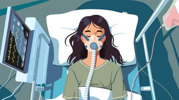
Congratulations to Dr. John Dick, Senior Scientist at UHN’s Princess Margaret Cancer Centre and University Professor at the University of Toronto, on his election to the National Academy of Sciences (NAS), one of the highest honours for a scientist.
On Tuesday, April 29, 2025, the NAS announced the election of 120 members and 30 international members in recognition of their continued achievements in original research. Membership in the NAS not only reflects extraordinary contributions to science but also entrusts inductees with shaping the future of research and policy at the highest levels.
With this recognition, Dr. Dick, known internationally as the first scientist to identify and characterize cancer stem cells, joins the membership of a scientific organization that includes an estimated 200 Nobel Prize winners.
Dr. Dick, who is also the Helga and Antonio De Gasperis Chair in Blood Cancer Stem Cell Research and a Professor of Molecular Genetics at the University of Toronto, is focused on understanding leukemic stem cells to develop better treatments and improve the quality of life for patients with leukemia. His work also earned him an induction into the Canadian Medical Hall of Fame in 2024.
Dr. Dick’s discovery of leukemic stem cells revealed that cancer cells are organized hierarchically, with only a small subset capable of self-renewal. This knowledge shifted research toward cancer stem cells, leading to more effective therapies targeting the cells responsible for the long-term spread of the disease.
The Manitoba-born Dr. Dick joins approximately 500 international researchers and 2,700 from the United States in a membership that is considered a mark of excellence in science.
Congratulations to Dr. Dick and all newly elected members!
The NAS, a private, non-profit society of distinguished scholars, was established by an Act of the United States Congress and signed by then-President Abraham Lincoln in 1863. Committed to furthering science, it aims to provide independent, objective advice on matters related to science and technology.
See here for the full announcement.
People living with metastatic breast cancer (MBC)—an advanced form of breast cancer where cancer has spread to other parts of the body—often face ongoing treatments and uncertainty. This raises complex questions about treatment decisions, symptom management, and future planning. To help address these challenges, researchers at The Institute for Education Research have developed an artificial intelligence (AI) chatbot that offers personalized, on-demand support and resources for people living with MBC.
The Artificial Intelligence Patient Librarian (AIPL) provides conversational guidance and tailored recommendations, suggesting helpful resources based on individual user questions.
To evaluate how well the chatbot works, researchers conducted a three-phase study:
● They first collaborated with MBC patients to adapt cancer education materials for use with the chatbot.
● Next, they trained the AI to scan and recommend information from over 100 reliable online resources chosen by expert patient librarians.
● They then tested the AIPL with 36 MBC patients through surveys and focus groups.
The participants found the chatbot easy to use and helpful for finding quick answers, especially for those who may be recently diagnosed. However, many participants were already well-informed and actively involved in their care. These users sought more in-depth medical information, emotional support, and guidance for managing relationships and planning for the future. Some also expressed interest in using the chatbot to connect with peers or explore alternative treatments and clinical trials.
This study shows how AI can help close gaps in cancer care by offering reliable and timely support for those navigating complex conditions. Future versions of the chatbot will aim to provide even more personalized help, including a digital companion that shares lived experiences. This could lead to more emotionally supportive care for people with advanced cancer. As this technology evolves, tools like the AIPL could transform how patients interact with their care—promoting connection, empowerment, and autonomy throughout their care journey.
The development and evaluation of the AIPL were conducted in collaboration with the M. Lau Breast Center at the Princess Margaret Cancer Centre, leveraging their expertise in patient education and support.
Dr. Yvonne Leung, the lead author of the study, is an Educational Investigator at The Institute for Education Research at UHN. Dr. Leung is also a Research Scientist at UHN’s de Souza Institute, an Adjunct Lecturer in the Department of Psychiatry at the University of Toronto, and a Teaching Assistant Professor at the North Eastern University, Toronto.
This work was supported by UHN Foundation and Pfizer Global Medical Grants.
Leung YW, So J, Sidhu A, Asokan V, Gancarz M, Gajjar VB, Patel A, Li JM, Kwok D, Nadler MB, Cuthbert D, Benard PL, Kumar V, Cheng T, Papadakos J, Papadakos T, Truong T, Lovas M, Wong J. The Extent to Which Artificial Intelligence Can Help Fulfill Metastatic Breast Cancer Patient Healthcare Needs: A Mixed-Methods Study. Curr Oncol. 2025 Mar 2. doi: 10.3390/curroncol32030145.
Acute myeloid leukemia (AML) is an aggressive blood cancer that arises when normal blood cell development goes awry. However, the disease does not follow a single pathway—each patient’s leukemia grows and develops in its own unique way. Understanding these differences has long been a challenge, but new research from the Princess Margaret Cancer Centre (PM) offers a clearer picture.
A team led by Dr. John Dick, Senior Scientist at PM, analyzed over 1.2 million leukemia cells from 318 AML patients, comparing them to a detailed atlas of healthy blood cells. This large-scale mapping effort revealed 12 key patterns of leukemia cell growth and development, showing how different genetic mutations drive the disease in distinct ways. Some leukemia cells resembled early-stage blood stem cells, while others mimicked more developed blood cells—findings that challenge traditional classifications of AML.
“These discoveries redefine how we see AML,” says Dr. Dick. “By understanding how leukemia cells develop, we can create treatments that precisely target a patient’s unique cancer.”
This research has immediate implications. Doctors could use these leukemia growth patterns to classify AML more accurately, leading to better treatment decisions. It also paves the way for personalized therapies that attack cancer at its root, based on its specific developmental pathway. Looking ahead, this map of leukemia’s hidden patterns could help researchers develop drugs that push leukemia cells back toward normal development—an approach known as differentiation therapy.
Dr. John Dick is a Senior Scientist and a Helga and Antonio De Gasperis Chair in Blood Cancer Stem Cell Research at the Princess Margaret Cancer Centre and Professor of Molecular Genetics at the University of Toronto. He is the senior author of the study.
Andy Zeng is an MD/PhD student in Dr. Dick’s lab at the University of Toronto and the first author of the study.
This work was supported by The Princess Margaret Cancer Foundation, the Canadian Cancer Society, the Ontario Institute for Cancer Research, the Canadian Institutes for Health Research, the University of Toronto’s Medicine by Design initiative with funding from the Canada First Research Excellence Fund, the Ontario Ministry of Health, a Canada Research Chair, and the Terry Fox Foundation.
Dr. John Dick has received research funding from BMS/Celgene and holds intellectual property licenses with Pfizer/Trillium Therapeutics. For a full disclosure of conflicts of interest, please review the publication.
Zeng AGX, Iacobucci I, Shah S, Mitchell A, Wong G, Bansal S, Chen D, Gao Q, Kim H, Kennedy JA, Arruda A, Minden MD, Haferlach T, Mullighan CG, Dick JE. Single-cell transcriptional atlas of human hematopoiesis reveals genetic and hierarchy-based determinants of aberrant AML differentiation. Blood Cancer Discov. 2025;OF1–OF18. doi:10.1158/2643-3230.BCD-24-0342.
Research conducted at UHN's research institutes spans the full spectrum of diseases and disciplines, including cancer, cardiovascular sciences, transplantation, neural and sensory sciences, musculoskeletal health, rehabilitation sciences, and community and population health.
Learn more about our institutes by clicking below: